Insights Into Autosomal Dominant Polycystic Kidney Disease From Genetic Studies
Mar 06, 2023
Abstract
Autosomal dominant polycystic kidney disease is the most common monogenic cause of ESKD. Genetic studies from patients and animal models have informed disease pathobiology. Strongly support a “threshold model” in which cyst formation is triggered by reduced functional polycystin dosage below a critical threshold within individual tubular epithelial cells due to germline and somatic PKD1 and PKD2 mutations, mutations of genes (e.g., SEC63, SEC61B, GANAB, PRKCSH, DNAJB11, ALG8, and ALG9) in the endoplasmic reticulum protein biosynthetic pathway, or somatic mosaicism. Genetic testing can potentially provide diagnostic and prognostic information on cystic kidney disease. However, mutation screening of PKD1 is challenging due to its large size and complexity, making it both costly and labor-intensive.
Moreover, conventional Sanger sequencing–based genetic testing is currently limited in elucidating the causes of atypical polycystic kidney disease, such as within-family disease discordance, atypical kidney imaging patterns, and discordant disease severity between total kidney volume and rate of eGFR decline. In addition, environmental factors, genetic modifiers, and somatic mosaicism also contribute to disease variability, further limiting prognostication by mutation class in individual patients. Recent innovations in next-generation sequencing are poised to transform and extend molecular diagnostics at reasonable costs. By comprehensive screening of multiple cystic diseases and modifier genes, targeted gene panel,whole-exome, or whole-genome sequencing is expected to improve diagnostic and prognostic accuracy to advance personalized medicine in autosomal dominant polycystic kidney disease.

FORE MORE Information:david.deng@wecistanche.com
Introduction
Autosomal dominant polycystic kidney disease (ADPKD) is a multisystem disorder characterized by the growth of numerous kidney cysts and expansion of kidney volume leading to ESKD in most patients. Hypertension, gross hematuria, cyst rupture and infection, kidney stones, and flank pain are common kidney complications. At the same time, extrarenal manifestations include liver and pancreatic cysts, vascular aneurysms, cardiac valve abnormalities, hernias, and diverticulosis. Estimating the prevalence of ADPKD has been challenging due to variable age-dependent penetrance and incomplete clinical ascertainment in the general population. Epidemiologic studies of clinically ascertained cases of ADPKD reported a point prevalence of 2.4–9.0 per 10,000. By contrast, a recent study of genomic sequencing in large populations yielded a minimal estimate of 1 per 1000. The identification of PKD1 in 1995 and PKD2 in 1996 facilitated the development of DNA sequence–based molecular diagnostics. Since then, driven by advancements in sequencing technology, our understanding of the complexities of the genetic basis of cystic kidney disease has evolved. Important messages underlying this review are that not all patients with multiple kidney cysts have ADPKD and that ADPKD and autosomal dominant polycystic liver disease (ADPLD) represent a phenotypic spectrum, with both diseases resulting from alterations in polycystin function. We discuss how genetic studies have informed our understanding of the pathobiology of ADPKD. We also discuss the current clinical indications for genetic testing in suspected ADPKD and its evolving role in elucidating the genetic causes of atypical polycystic kidney disease (PKD). A glossary of terms used in this review is provided in Box 1.
Genetic Studies Informed Disease Mechanisms in ADPKD
Mutations of two genes, PKD1 and PKD2 (which encode two integral membrane proteins, polycystin-1, and polycystin-2 [or TRPP2], respectively), are responsible for most genetically resolved cases of ADPKD. Polycystin-1 is a large protein with functional features suggestive of a receptor of unknown function, whereas polycystin-2 is a nonspecific cation channel; both interact through their cytoplasmic tails to modulate a novel signaling pathway in the primary cilia. Variable disease expression is a notable feature of ADPKD. Even though the inherited germline defect is present in all cells, cysts form in,5% of tubules, and cystic dilation is focal within each tubule. These observations, coupled with the discovery of somatic PKD1 or PKD2 mutations in kidney and liver cysts of patients with ADPKD, led to the hypothesis of a recessive cellular mechanism for cystogenesis in which the inactivation of both copies of either PKD1 or PKD2 in an individual tubular epithelial cell
is required to initiate cyst formation. However, this hypothesis was initially challenged because of a low proportion of somatic PKD1 mutations identified in the cysts of patients with germline PKD1 mutations, albeit only the nonduplicated region of PKD1 was screened in earlier studies. Using locus-specific PCR and next-generation sequencing (NGS), a recent study comprehensively screened and identified private somatic PKD1 and PKD2 mutations in 90% of 128 cysts from nine patients, providing the most definitive evidence in support of the above hypothesis. However, complete inactivation of both copies of PKD1 or PKD2 is not necessary for cyst initiation as studies f hypomorphic mutant mouse models showed cyst development with low levels (approximately 20%) of polycystin-1. Currently, the “threshold” model of cystogenesis provides the best explanation of human and animal studies to date. Accordingly, reduced functional polycystin-1 dosage below a critical threshold (i.e., approximately 20%–30%) within tubular epithelial cells due to germline and somatic PKD1 or PKD2 mutations, the type (i.e., protein-truncating versus nontruncating) of mutation, and local stochastic factors appear to trigger individual cyst formation. However, timing also seems to be important: Pkd1 inactivation before 13 days after birth in a mouse model resulted in severe cystic disease within 3 weeks; by contrast, Pkd1 inactivation after 14 days of age in the same model resulted in an indolent course with cyst development only after 5 months. Moreover, cystic kidney disease in the late induction model was exacerbated by kidney injuries, such as ischemia-reperfusion and tubular obstruction from crystal deposition (also known as the “third-hit model”). Collectively, these findings suggest that additional factors beyond the “second hits” contribute to the expansion of dilated tubules into macrocysts. Intriguingly, a recent study of Pkd1 mutant mice suggests that local factors arising from individual cysts may promote adjacent dilated tubules to evolve into frank cysts in clusters (also known as the “snowball effect”).
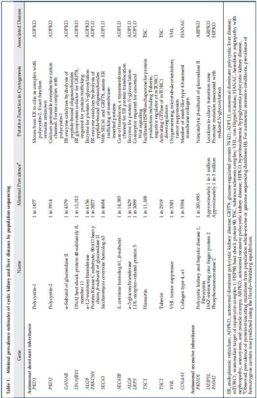
Recent genetic studies have identified a novel mechanism by which mutations in multiple genes encoding proteins functioning in the endoplasmic reticulum (ER) protein biosynthetic pathway cause ADPLD by modulating polycystin-1 dosage (Table 1). Specifically, translocation proteins encoded by SEC63 and SEC61B are required for the entry of nascent proteins into the ER, whereas the proteins encoded by ALG8, ALG9, and PMM2 are required in the ER for N-glycosylation of nascent proteins. Glucosidase II, composed of an a-subunit encoded by GANAB and a b-subunit encoded by PRKCSH, removes glucose molecules from the nascent proteins after quality control by the calnexin/calreticulin cycle before they are exported to the Golgi complex (Figure 1). Additionally, a cofactor of the binding-Ig protein encoded by DNAJB11 functions as a chaperone in the ER for control of protein folding, trafficking, and degradation, including polycystin-1 and polycystin-2. Mutations of any of the above genes can reduce the functional polycystin-1 dosage by impairing its post-translational modification and trafficking to the cell membrane and thus, can modify cystic disease severity in the setting of ADPKD.

Figure 1. | Schematic representation of endoplasmic reticulum maturation and N-glycosylation of nascent polycystin-1 (PC-1) and polycystin-2 (PC-2). Mutations in PKD1 and PKD2 cause autosomal dominant polycystic kidney disease (ADPKD); mutations in SEC61B, SEC63, ALG8, GANAB, and PRKCSH cause autosomal dominant polycystic liver disease; mutations in PKHD1 cause autosomal recessive polycystic kidney disease; and mutations in ALG9 and GANAB cause atypical ADPKD. All conditions have phenotypic overlap and include some degree of kidney and liver cysts, and they are related to alterations in the functional dosage of mature PC-1/PC-2 complex.
The mechanisms by which reduced polycystin signaling in the primary cilia of tubular epithelial cells leads to cystic disease remain incompletely understood. Experimental therapies targeting increased cAMP, activation of the mammalian target of rapamycin complex 1 (mTORC1), and reduced 5ʹ-AMP–activated protein kinase signaling have been shown to slow cystic disease progression (Figure 2). Tolvaptan, a vasopressin receptor 2 antagonists, has been successful in slowing the progression of ADPKD in humans by reducing cAMP signaling. Of interest, a recent study of murine PKD models showed that ablation of primary cilia by inactivation of a ciliary gene (either Kif3 or Ift20) resulted in mild disease, whereas inactivation of either Pkd1 or Pkd2 resulted in severe disease. Unexpectedly, ablation of the primary cilia in addition to the inactivation of Pkd1 or Pkd2 in these models resulted in attenuated kidney disease compared with Pkd1 or Pkd2 inactivation alone. Taken together, these data implicate the existence of a yet unidentified cilia-based signaling pathway that interacts with the polycystin complex to modulate cyst disease severity and loss of cilia function seems to be protective in the context of ADPKD.

Evolution of Mutation Screening Technologies in ADPKD
PKD1 mutation screening has been challenging due to its large size, complexity, and high GC content. The gene comprises 46 exons and encodes a 12,912-bp transcript, spanning a 50-kb genomic region. Its first 33 exons are duplicated in six pseudogenes with approximately 98% DNA sequence identity. The high level of DNA sequence identity with the pseudogenes creates the possibility for both false positive and negative genotype calls because a pseudogene mutation can be incorrectly called as present in PKD1, and a PKD1 mutation can be missed if the signal is overwhelmed by the normal sequence in the pseudogenes when DNA capture assay is used. The first protocol for a comprehensive PKD1 mutation screen exploited rare mismatches between the duplicated region of PKD1 and its pseudogenes to generate locus-specific templates for subsequent nested sequencing reactions. Five long-range PCRs are required to generate PKD1-specific amplicons from the duplicated region followed by 65 nested PCRs to screen the entire gene. This protocol provides a robust safeguard against spurious genomic amplification from the PKD1 pseudogenes and yielded a high diagnostic rate of approximately 80%–90% in selected clinical cohorts, but it is labor-intensive and costly.

Figure 2. | Insights into the pathobiology of ADPKD from human and animal genetic studies. AMPK, 5ʹ-AMP-activated protein kinase; ER, endoplasmic reticulum; ERK, extracellular signal–regulated kinase; JAK-STAT, Janus kinase-signal transducer and activator of transcription signaling pathway; mTOR, mammalian target of rapamycin.
A subsequent protocol generated long-range locus-specific PCR amplicons for both genes (i.e., eight for PKD1 and six for PKD2) and multiplexed them from individual patient samples as bar-coded libraries for high-throughput sequencing. This latter approach significantly reduces laboratory workload and costs and is currently used by several research laboratories; however, it still requires considerable technical expertise. More recently, targeted exome sequencing by customized gene panels using DNA capture or whole genome sequencing has been applied for PKD1 and PKD2 mutation screening, with promising results and .95% accuracy for previously resolved cases; however, sophisticated bioinformatics analyses are required.
Mutation Class Predicts Average Kidney Disease Severity in ADPKD
Mutation screening of patients with ADPKD enriched with high-risk clinical features for progression reports that 75% of cases carried PKD1 mutations, approximately 15% carried PKD2 mutations, and at least 10% of cases had no mutation detected. By contrast, mutation screening of patients ascertained with normal or near-normal kidney function reported that 60% of cases carried PKD1 mutations, 25% carried PKD2 mutations, and 15% had no mutation detected. Over 1250 PKD1 mutations and 200 PKD2 mutations have been archived in the Mayo Polycystic Kidney Disease database, and no single mutation accounts for .2% of cases. Multiple studies have confirmed a strong correlation between mutation class and kidney disease severity: on average, protein-truncating PKD1 mutations (i.e., frameshift, nonsense, and canonical splice site mutations and large deletions) are associated with the most severe disease(mean age of ESKD: 50–55 years), followed by nontruncating PKD1 mutations (i.e., missense and in-frame insertions/deletions) and PKD2 mutations (mean age of ESKD: approximately 75–80 years). However, significant disease variability within families of affected relatives with the same main effect mutation is well documented and strongly suggests a modifier effect. Additionally, in the absence of a robust in vitro assay, there is uncertainty for the assignment of pathogenicity in nontruncating variants. The American College of Medical Geneticists has developed guidelines for the interpretation of rare sequence variants that includes the evaluation of allele frequency in population databases, presence as pathogenic in disease databases, in vitro or in vivo functional characterization, bioinformatic evaluation, and cosegregation with the disease in multiple affected or absence in unaffected family members. Variants are then reported as “pathogenic,” “likely pathogenic,” “variant of uncertain significance,” or “benign.” However, the cumulative population frequency of “likely pathogenic” variants in PKD1 and PKD2 exceeds the epidemiologic estimates of the prevalence of ADPKD, calling the penetrance of some of these variants into question.
Genetic Causes of PKD beyond PKD1 and PKD2
Despite comprehensive screening, 10%–15% of patients suspicious of ADPKD have no mutation detected in either PKD1 or PKD2. Using whole-exome sequencing, mutations have been identified in several additional rare cystic disease genes in patients who were originally labeled as “no mutation detected”. As described above, multiple genes (i.e., ALG8, ALG9, GANAB, PRKCSH, SEC61B, and SEC63) encoding proteins that function in the ER biosynthetic pathway have been shown to cause ADPLD, and a clinical picture that overlaps with ADPKD. Autosomal recessive mutations in PMM2 cause a devastating pediatric multisystem disorder, but a rare promoter mutation that reduced PMM2 expression has been identified to cause childhood-onset hyperinsulinemic hypoglycemia and polycystic kidneys in fifteen families. Mutations in DNAJB11 have been shown to cause atypical PKD with small kidney cysts, small or normal kidney size, and late-onset kidney failure associated with tubular atrophy and interstitial fibrosis—clinical features of both ADPKD and autosomal dominant tubulointerstitial kidney disease (ADTKD). This discordance of kidney size with function is highly unusual for ADPKD but is also seen in ADPLD associated with ALG9 mutations.

Somatic mosaicism is another important cause of ADPKD in patients with no mutation detected. Mosaicism refers to the presence of two genetically distinct cell populations within one individual resulting from a somatic mutation during embryogenesis. Depending on the cell type and developmental stage when the mutation occurs, three clinical syndromes can arise germline mosaicism, involving the germ cells only; somatic mosaicism, involving the body cells only; and gonadal and somatic mosaicism, involving both the germline and somatic cells. The presence of de novo PKD with atypical kidney imaging patterns (i.e., asymmetric, unilateral, lopsided patterns) is a clinical finding suspicious of somatic mosaicism. The diagnosis of mosaicism is challenging due to the variable involvement of the affected cells resulting in a low mutation signal-to-noise ratio, and it is frequently missed by Sanger sequencing. However, approximately 10% of genetically unresolved cases from three clinically ascertained cohorts were found to harbor somatic mosaicism by NGS. All identified somatic mutations were found in PKD1 sequenced from peripheral blood DNA at variant allele fractions between 5% and 20%. Future studies using “molecular barcoding” of template DNA from different tissues (e.g., buccal mucosa and urinary epithelia) may improve the detection rate of mosaic cases with even lower variant allele fractions (i.e.,,2% of reads).
Current Indications for Genetic Testing in ADPKD
For most at-risk subjects with a positive family history, the diagnosis of ADPKD can be confirmed by ultrasound or magnetic resonance imaging using well-validated, age-dependent criteria based on kidney cyst counts (Table 2). However, disease exclusion with complete certainty may not be possible in at-risk subjects younger than 40 years of age by ultrasound. By contrast, with higher resolution for detecting small cysts, magnetic resonance imaging can be used for disease exclusion with high certainty. Clinical genetic testing for ADPKD is currently indicated in cases where there is doubt regarding the diagnosis (i.e., lack of family history or equivocal imaging findings) or where there is a need for disease exclusion with high certainty at an early age, such as in the case of the workup for a potential living kidney donor or prenatal and preimplantation genetic diagnosis (Table 3). Early exclusion of ADPKD in an at-risk subject can be performed by genetic testing for the specific familial mutation. We do not recommend screening minors at risk of ADPKD beyond BP monitoring because disease-modifying treatment is lacking in this population, and obtaining a diagnosis in a presymptomatic state can cause psychological stress. All subjects undergoing genetic testing for ADPKD should be informed of the potential ramifications of genetic testing, which vary from country to country, and should receive pre-and post-test counseling by a genetic counselor.

Evolving Indications for Genetic Testing in ADPKD
Advances in NGS are poised to revolutionize genetic testing in ADPKD by providing simultaneous mutation screening of multiple cystic diseases and potential modifier genes with high accuracy and reasonable costs (28). As such, we expect that several new indications will evolve with advancing high-throughput sequencing methodologies targeting atypical forms of PKD (Figure 3, Table 3); they include (1) early and severe disease, (2) marked intrafamilial disease variability, (3) no apparent family history, (4) atypical kidney imaging, and (5) syndromic presentation. The cumulative prevalence of these atypical scenarios may be as high as one-third of patients with ADPKD. We suggest that patients or families displaying one of these scenarios should be referred to specialized centers for further testing.
(1) Early and Severe Disease
Severe PKD presenting in utero or early childhood is a well-described clinical entity. For example, the contiguous PKD1 and TSC2 deletion is a rare syndrome associated with enlarged cystic kidneys, variable signs of tuberous sclerosis complex (TSC), and, typically, ESKD by teenage years More recent studies have demonstrated genetic complexity underpinning some of these severe cases, including compound heterozygosity (e.g., due to one protein-truncating PKD1 mutation in trans with a second nontruncating PKD1 mutation) or digenic disease (e.g., due to one PKD1 mutation and a second mutation in another cystic disease gene, such as PKD2, COL4A1, or HNF1B). Homozygous loss-of-function PKD1 or PKD2 mutations are believed to be embryonically lethal in humans. Although rare, the identification of families with bilineal ADPKD has important implications for genetic counseling. Although it may be suggested from the family history, this is often not the case because one affected parent may have a mild form of ADPKD (i.e., due to a nontruncating PKD1 or a PKD2 mutation). Clinical clues for potential bilineal disease in ADPKD may include marked kidney disease discordance between family members and a high disease segregation ratio affecting approximately 75% of children in large pedigrees, as opposed to the 50% expected in an autosomal dominant condition, due to the two independently segregating mutations.

Figure 3. | Clinical scenarios of atypical ADPKD presentations and potential genetic explanations.
(2) Marked Intrafamilial Disease Variability
Marked kidney disease discordance (i.e., by total kidney volume or eGFR, adjusted for age) in an affected relative pair is relatively common, affecting at least 12% of families with ADPKD, and may result from the coincidence of second kidney disease (e.g., diabetes or GN) in the more severely affected member or the presence of an unusual genetic underpinning (i.e., somatic mosaicism or genetic modifiers, including digenic disease as discussed above). Digenic or biallelic disease associated with different genic or allelic effects in patients with ADPKD has been reported and strongly supports the “threshold model” of cystogenesis in which cellular functional polycystin dosage inversely correlates with disease severity (Figure 4). In addition, comorbidities (such as hypertension and obesity) and environmental factors (such as smoking and water intake) can also contribute to disease discordance within families.
(3)No Apparent Family History
Up to 28% of patients suspected to have ADPKD report no apparent family history. In this setting, genetic testing may be indicated, and the differential diagnosis needs to broaden to include other genetic and nongenetic causes of cystic kidney disease, especially in cases with atypical or syndromic features. Other differential diagnoses include a de novo mutation, unavailability of parental medical records, somatic or germline mosaicism, or mild ADPKD unrecognized in an affected parent. In case of suspected somatic mosaicism, screening with high read depth may also be helpful to resolve the genetic cause.

Figure 4. | Effects of digenic disease on functional polycystin dosage and cystic disease severity. hp, hypomorphic variant.
(4) Atypical Kidney Imaging
Atypical kidney imaging patterns (i.e., Mayo Clinic Imaging Class 2) are present in up to 16% of patients suspected to have ADPKD. Patients without a family history of ADPKD who displayed unilateral, asymmetric, segmental, or lopsided cystic disease are suspicious of somatic mosaicism. By contrast, patients with positive family history and atypical kidney imaging tend to display mild cystic disease associated with nontruncating PKD1 or PKD2 mutations. Patients with moderate to advanced kidney failure but kidney imaging showing mild cystic disease without kidney enlargement should raise the suspicion of a second kidney disease, such as diabetic nephropathy, GN, or another cystic disease. In this setting, the differential diagnosis should also include PKD due to mutations in DNAJB11 or ALG9, thin basement membrane disease (due to a heterozygous COL4A3 or COL4A4 mutation), or apoL1 kidney disease in a Black patient with proteinuria.
(5) Syndromic Presentation
Table 1 provides a list of genes that can lead to cystic kidney diseases when mutated. Of note, syndromic manifestations in these disorders may often provide clues
to their diagnosis. For example, autosomal recessive PKD may present in young adulthood with congenital hepatic fibrosis or Caroli syndrome. The presence of hamartomas in the targeted organs usually allows for an unambiguous diagnosis of TSC. However, differentiation of TSC (including the PKD1-TSC2 contiguous gene deletion syndrome) from ADPKD may be difficult in the presence of somatic mosaicism. Additional cystic kidney diseases not associated with kidney renal enlargements, such as those caused by mutations in DNAJB11 or ALG9, should be considered. ADTKD includes several disorders that are characterized by progressive CKD associated with low-grade proteinuria and bland urinalysis. Small kidney cysts may develop in the late clinical course, and additional clinical features may help to differentiate these conditions. For example, the presence of gout and hyperuricemia is suggestive of ADTKD associated with uromodulin mutations, whereas the presence of maturity-onset diabetes of the young and/or genitourinary tract malformation is suggestive of ADTKD associated with HNF1B mutations. Other rare forms of PKD with distinctive syndromic features include von Hippel– Lindau disease; nephronophthisis; X-linked dominant orofaciodigital syndrome; hereditary angiopathy, nephropathy, aneurysms, and muscle cramps syndrome; and hyperinsulinemia hypoglycemia with PKD. By comprehensively screening a list of known and potential cystic disease genes, genetic testing with high-throughput sequencing is expected to improve diagnostic accuracy in patients with cystic kidney disease.
Genetic studies from patients and animal models have informed disease pathobiology in ADPKD. We now understand that reduced functional polycystin dosage below a critical threshold, through both genetic and nongenetic mechanisms, triggers cyst formation within individual tubular epithelial cells. However, the exact molecular mechanisms underlying cyst growth and disease progression remain incomplete. NGS-based genetic testing is expected to improve the diagnostic accuracy of cystic kidney disease and may also provide prognostic information for ADPKD. Conventional Sanger sequencing-based genetic testing is limited in elucidating the causes of atypical PKD presenting in clinical scenarios, such as within-family disease discordance, atypical kidney imaging patterns, discordant disease severity between eGFR decline and total kidney volume, and another syndromic cystic kidney disease. Innovations in high-throughput sequencing will transform and extend molecular diagnostics in ADPKD. Through comprehensive screening of multiple cystic diseases and modifier genes and improved detection of somatic mosaicism, targeted gene panel, whole-exome, or whole-genome sequencing is expected to improve diagnostic and prognostic accuracy to advance personalized medicine in ADPKD.

Box 1.Atypical Imaging PatternUnilateral, segmental, asymmetric, or lopsided kidney cyst distributions that differ from the classic symmetric distribution observed in autosomal dominant polycystic kidney disease. Bilineal DiseaseDescribes when a mutation has been inherited from both parental lineages. Includes compound heterozygosity, where both mutations are in the same gene, or digenic inheritance, where the mutation from each parent is in a different gene. De Novo MutationA new genetic variant is present for the first time in a family. Germ CellsA sperm or the unfertilized egg contains one copy of each autosomal chromosome and one sex chromosome. High-Throughput SequencingAlso known as next-generation sequencing, it represents a group of massively parallel sequencing technologies that allow for the sequencing of hundreds of millions of reads per experiment. Used in gene panel, whole exome, and whole-genome sequencing. Lopsided Imaging PatternAn atypical kidney cyst imaging pattern whereby fifteen or fewer very large cysts account for .50% of the total kidney volume. Minor Allele FrequencyThe prevalence of the less common allele of a genetic variation in the population. Molecular Bar CodingThe addition of a short segment of DNA to label a DNA fragment. After sequencing, the label can be used to determine which source the fragment came from, be it from different people, tissue types, or rounds of PCR. Polygenic InheritanceThe cumulative result of many small effect variants throughout the genome. Read DepthAlso known as coverage, it is the average number of times that each base is read per sequencing run. Greater read depth increases confidence in the genotype call at each nucleotide and increases the likelihood of detection of a variant with low variant allele frequency (a somatic variant). Somatic MutationA genetic alteration occurs to a cell at some point after fertilization that will be passed down to progeny cells during cellular replication. Variant Allele Frequency (or Fraction)The proportion of reads that contain an alternate allele from a single person. In a heterozygous person, a variant allele frequency of 50% is expected, but deviations can occur due to variable PCR or sequencing efficiency. In somatic mosaicism, variant allele frequency can be very low (i.e.,,2% of reads). |
Disclosures
M.B. Lanktree has received compensation for participation as a speaker and an advisory board member for Otsuka Pharmaceuticals. He also received grants from the Kidney Foundation of Canada during the conduct of the study. Y. Pei has received compensation for participation in advisory boards for Otsuka, Reata Pharmaceuticals, and Sanofi-Genzyme. All remaining authors have nothing to disclose.
Funding
This work was supported in part by the Canadian Institutes of Health Research Strategy for Patient-Oriented Research Program Grant in Chronic Kidney Disease Can-SOLVE CKD Network Program. M.B. Lanktree is a new investigator in the Kidney Research Scientist Core Education and National Training (KRESCENT) program funded by the Canadian Institutes of Health Research, the Kidney Foundation of Canada, and the Canadian Society of Nephrology.
FORE MORE Information:david.deng@wecistanche.com